Cell Counter

Bright Field Cell Counter:
Three models are available which uses the bright field imaging for cell counting and assay.
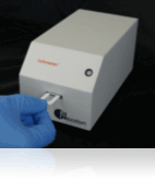
The Cellometer Mini is the most basic model of Nexcelom which utilizes bright field imaging and pattern-recognition software to quickly and accurately identify and count individual live cells and dead cells stained with Trypan Blue. It helps to measure the cell size, viability and concentration of cell lines and few cell based assay in less than 10 sec.
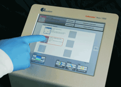
The Cellometer Auto 1000 is similar to Cellometer Mini except that it has onboard display with in built software for analysis.
The Cellometer Auto T4 is similar to Auto 1000 but is suitable for GMP facility as it has the provision for GMP software for analysis.
Fluorescent Cell Counters
These are the cell counter that works with fluorescence dye for better assay and analysis.
The Auto 2000 utilizes bright field imaging and dual-fluorescence imaging to quickly and accurately identify and count individual cells. Cell count, concentration, diameter, and % viability are automatically calculated and reported. Dual-colour fluorescence allows for staining of live and dead nucleated cells, generating accurate viability results even in the presence of debris, platelets, and red blood cells. Accurate analysis of both ‘messy’ and ‘clean’ samples enables the Auto 2000 to evaluate samples at a variety of points throughout sample processing – from initial collection to separation, to cryopreservation
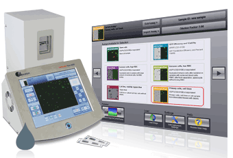
Cellometer K2 is again similar to Auto 2000 and uses double Fluorescence for analysis of Hepatocytes, stem cells, splenocytes, tumor suspension and other complex primary cells. Has ability to export to FCS Express for cell population analysis. Optional GMP/GLP software is available.
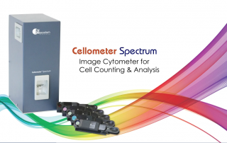
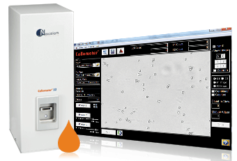
Cellometer Spectrum cell counter and Image Cytometer is a all in one system and advance version instrument. It is customizable cell counting and cytometry instrument that provides flow-like data with predesigned templates. Quickly plot cell population data as a histogram, scatter plot, dot plot or contour plot using FCS express. The filter sets are user-changeable, allowing you to customize the instrument for specific assays/reagents of interest. It allows to have fluid like data without having inconvenience of regular washing of fluid channel after every assay.
Plate based Image Cytomere:
These are the advance version of cell counter as well as image cytometery which is used when analysis of sample is needed in large number or in different format of wells.
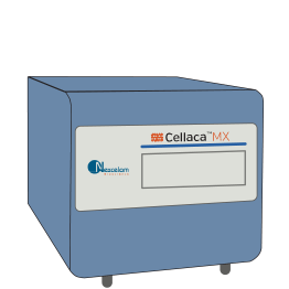
Cellaca MX is a high-throughput Automated Cell Counter that can count up to 24 samples in 48 seconds using trypan blue or in 2.5 minutes with fluorescence. One can run batch samples in 24 well microplates for cell count and viability. Cellaca’s ability to consistently image, de cluster, and report cell viability and concentration make it a versatile instrument that can be used in multiple workflows. The ability to add additional automation with robotic integration can further increase productivity.
Celigo is a bench-top, micro-well plate based, bright field and fluorescent imaging system designed for live cell analysis. The best-in-class bright field imaging capability, in combination with 4 fluorescence channels, provide high speed, fully automated imaging and quantification of suspension, adherence cells, tumor spheroids, iPSC and cancer stem cell colonies within 6, 12, 24, 48, 96, 384, 1536-well plates, T flasks, and slides. Proprietary optics and scanning system enable fast imaging of entire well while maintaining consistent illumination and contrast out to the well edge, for accurate identification of all cells within each well. With an intuitive software interface and optional integration into automation platforms, the Celigo provides labs with increased capabilities.
Yeast Cell Counter:
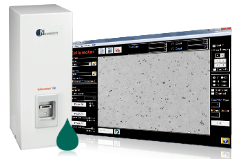
The Cellometer X2 Image Cytometer is specifically optimized for simple, 1-step determination of yeast concentration and viability and makes it suitable for research laboratories and small and large breweries looking to automate their fermentation monitoring. Bright field images can be viewed to confirm cell morphology. Counted bright field images show counted cells within cell clumps. Fluorescent images show dead cells for confirmation of viability results. Analysis of both bright field and fluorescent images from each sample makes 1-step concentration & viability determination possible.